


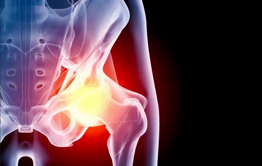
Hip arthrosis (coxarthrosis) is a chronic pathology accompanied by the gradual destruction of cartilage tissue in the affected area and then the involvement of adjacent structures in the process. The disease requires long-term treatment, and in severe cases, the only way to regain mobility is joint replacement.
general information
Coxarthrosis belongs to the group of degenerative diseases. It gradually begins with microscopic changes in the structure of the cartilage. Increased stress, inflammatory diseases, blood supply disorders lead to structural changes and thinning of cartilage tissue, thereby deforming the contours of the joint area. As a result, the distribution of the load on the contact surfaces of the bones changes and the zones of maximum pressure begin to wear faster. This initiates a complete cascade of pathological reactions:
- the appearance of microcracks and compression areas in cartilage tissue;
- decreased smoothness of joint surfaces;
- cartilage overgrowth at the site of thinning and replaced with bone tissue;
- the appearance of osteophytes (bone growths) along the edge of the joint;
- thickening and loss of elasticity of the joint capsule;
- hardening and reduced strength of the strips;
- changes in the composition of the synovial fluid (natural lubrication within the joint);
- narrowing of the common space;
- fusion of all elements of the joint (ankylosis).
Without treatment, coxarthrosis inevitably becomes the cause of immobility and disability.
Cause
Depending on the causes of the disease, a distinction is made between primary and secondary coxarthrosis. In the first case it occurs on its own, for example in the background of an inherited predisposition, in the second it is provoked by other diseases or injuries. In most cases, the process of cartilage degeneration is due to a combination of several factors. This may be due to:
- congenital dislocation of the hip;
- flat legs, scoliosis and other orthopedic problems;
- Legg-Calve-Perthes disease;
- arthritis (iris inflammation), regardless of origin;
- injuries and microtraumas of the hip joint, overweight, professional sports, etc. background;
- hip dysplasia;
- metabolic disorders;
- endocrine diseases (especially diabetes);
- violation of the blood supply to the lower extremities;
- frequent stress;
- inheritance (coxarthrosis of parents or other close relatives significantly increases the risk of developing a child)
- congenital pathologies of the connective tissue and autoimmune diseases (joint hypermobility, rheumatoid arthritis, systemic lupus erythematosus, etc. );
- they have undergone joint operations.
Age is an important predisposing factor. According to statistics, after 45 years, the likelihood of developing coxarthrosis increases significantly.
Symptoms
The main symptoms of coxarthrosis of the hip joint do not depend on the cause of the development. Most patients note:
- restriction of movement: one of the earliest symptoms is due to thinning of the cartilage layer and increased friction on the joint surfaces of the bones; the emergence of cartilage growth in the future will exacerbate the problem;
- pain: friction between the bones deprived of the cartilage layer, the gradual involvement of all elements of the joint in the degenerative process, a decrease in the blood supply to the tissues causes increased pain sensations as the disease progresses; the pain is of a shooting nature and often worsens towards the end of the day;
- muscle cramps leading to increased pain symptoms and limited range of motion in the joint;
- decrease in leg length: this symptom appears in the later stages of the disease due to narrowing of the joint space and constant friction due to the gradual grinding of the bone head; the difference between the legs must not exceed 2 cm;
- lameness: accompanied by severe pain and limited mobility and shortening of the legs; an unfavorable sign indicating severe damage to the joint device.
Platoon
During development, coxarthrosis goes through several stages that depend on the degree of tissue damage.
- 1 degree. During this time, the patient experiences mild painful pain in the joint that occurs after intense or prolonged physical activity and resolves rapidly after rest. As a general rule, discomfort occurs exactly in the area of the hip joint, but in some cases extends to the hip or knee. The gait does not change, the leg movements are fully retained. Specific changes are observed on the roentgenogram: subchondral sclerosis.
- Grade 2. The pain becomes more severe, occurs after exertion, spreads to the entire thigh and groin. Mild lameness may occur after exertion. There are difficulties in abducting a foot. X-ray shows a significant decrease in the distance between the bones (by 50% or more), deformity of the femoral head, and marked bone growth.
- 3 degrees. The pains become constant, walking without reeds becomes impossible. During movement, the patient noticeably leans towards the painful side, which further increases the load on the joint. The range of motion decreases, the muscles of the legs and buttocks wither. The affected limb is shortened. X-ray reveals significant joint deformity, changes in the contour of the femoral head, and a large number of osteophytes.
- 4 degrees. The pain becomes stronger and does not stop for a minute, the patient loses the ability to move independently. X-rays show complete destruction of the articular cartilage as well as signs of fusion of the bones (ankylosis). Coping with the disease at this stage is only possible with surgery.
Diagnostics
An orthopedic traumatologist is responsible for identifying symptoms and selecting treatment. Use the following to diagnose and quantify the disease:
- survey: listening to patients' complaints, identifying risk factors (trauma, illness, heredity, etc. );
- examination: assessment of limb mobility, identification of areas of greatest pain;
- X-ray: X-ray makes it possible to assess the condition of bones and cartilage, the size of the joint space, the presence and location of bone growths; the examination is supplemented by CT (computed tomography) in order to have a more thorough overview of the necessary details;
- laboratory diagnostics: a general blood test allows the identification of biochemical signs of the inflammatory process - to note some risk factors such as uric acid levels;
- MRI (magnetic resonance imaging): allows you to assess not only the condition of bones and cartilage, but also the condition of soft tissues: bones, ligaments, muscles, joint capsules, etc.
- joint defect.
If it is necessary to perform differential diagnosis with other diseases as well as to assess concomitant pathologies, additional examinations, instrumental examinations, and consultation with narrow specialists are prescribed.
Coxarthrosis treatment
Treatment of hip coxarthrosis depends on its stage and severity of symptoms. Pathology requires an integrated approach using different methods:
- drug treatment;
- non-pharmacological treatment (physiotherapy, training therapy);
- surgery;
- lifestyle improvement and diet.
Drug treatment
The purpose of drugs prescribed for hip arthrosis:
- removal of pain syndrome;
- restoring or at least slowing down cartilage tissue;
- improving the blood supply and nutrition of the affected area;
- treatment of concomitant pathologies.
Analgesics are used in the form of tablets, intramuscular and intraarticular injections and topical agents: creams, ointments, patches. In the early stages of the disease, nonsteroidal anti-inflammatory drugs are sufficient for most patients. In case of severe pain syndrome, hormonal agents are used. The introduction of analgesics directly into the joint capsule has a good effect.
If the course of the disease is accompanied by muscle cramps, muscle relaxants are used. It is used in combination with other painkillers.
The use of analgesics should be limited in time and dose so as not to cause further damage to cartilage and other side effects (especially gastritis and gastric ulcer).
Chondroprotectors are drugs that help restore cartilage tissue. They are only effective with long-term regular use, combined with other treatments, lifestyles, and dietary adjustments. Drugs that improve blood microcirculation help increase their effect. Warming ointments are prescribed for a similar purpose. Only the doctor is involved in the selection of dosage and treatment.
Drug-free treatment
This category includes a variety of physiotherapy and hand techniques as well as physiotherapy exercises. They help improve microcirculation and restore movement of the damaged joint. Depending on the situation, your doctor will prescribe:
- shock wave therapy;
- magnetotherapy;
- electromyostimulation;
- various types of electrophoresis and phonophoresis (including administration of anesthetic drugs);
- mechanotherapy;
- massage and exercise.
Surgery
Once the disease has reached stages 3-4 of development, medications and physiotherapy will only alleviate the patient’s condition but will not restore full mobility. In this case, arthroplasty is indicated, i. complete or partial replacement of the damaged joint with a titanium prosthesis.
If there are signs, an easier version of the procedure is performed: sanding the contact areas of the bones and covering them with special smooth implants that make it easier to slide.
Prevention
Lifestyle can significantly reduce the risk of developing coxarthrosis as well as the rate of progression. Strict adherence to the rules is important:
- lead an active lifestyle: swimming in the pool, walking, cycling - physical activity at the amateur level without competing for records, improves blood supply and inhibits the processes of joint degeneration;
- normalizes body weight to reduce leg strain;
- eliminate injuries, hypothermia, and workplace risk factors (vibration, weightlifting, standing work);
- treat all diseases in a timely manner, including those not directly related to the musculoskeletal system;
- correct posture disorders in a timely manner, wear comfortable shoes.
Diet
With nutritional correction, the patient can not only reduce their weight, but also reduce inflammatory reactions, salt deposits in the tissues, and metabolic disorders. We recommend that you stick to a balanced menu that includes enough but not excessive amounts of carbs, protein, and fat, as well as vitamins and minerals. Particular attention should be paid to unsaturated fats (olive and flaxseed oil), omega-3 acids (found in excess in fish), collagen (jelly meat, aspic). It is recommended to minimize fast carbohydrates, alcohol, strong coffee, artificial flavor products, preservatives and flavor enhancers.
Consequences and complications
Coxarthrosis is a common cause of disability in the elderly. Without proper treatment, the pathology inevitably leads to complete disability, especially in bilateral lesions. Pain and restraint do not allow you to work and take care of yourself, so it is important to treat your treatment in a timely manner.